Image

FAREWELL!!!!!

FAREWELL!!!!!

Well folks we have come to end….. 😦 ….. We have learnt alot and would like to take this opportunity to thank our lecturer. Sir you have truly been an inspiration. You have encouraged us , and supported us in every way and you have always been a great help. You made complicated things seem easy and with your personality combined, this course was truly enjoyed, and we would like to take this moment to express our appreciation for you being with us. To our fellow students we had fun and will all put our best foot forward and strive for success. GOOD LUCK IN EXAMS!!!!!!!!! And hope to see you all next Semester…. 😀

Nucleotides and Nucleic Acids

Crick’s Central Dogma Genes are expressed by replication, translation and transcription of DNA. The DNA is replicated and the genetic information is translated in the form of RNA. The RNA transcripts the genetic information in the form of a proteinwhich then expresses the gene.


. Image

Image 107


Nucleotides are made up of a base, pentose sugar and a phosphate group. The base and the sugar make up the nucleoside. When the phosphate group is added is then a nucleotide. Nucleotides contain oxygen, carbon, phosphorus, nitrogen and hydrogen. The bases attached to the nucleotide are either pyrimidine or purines.

Image 108  

Pyrimidines are thymine, uracil and cytosine. Uracil is found only in and RNA while thymine and cytosine is found in both RNA and DNA. Uracil and thymine are similar in structure so uracil replaces thymine in RNA.

Image

Image 109

Purines are guanine and adenosine. Both are found in DNA and RNA.   Erwin Chargaff discovered that there were equal ratios of adenine to thymine and guanine to cytosine in most DNA of different species. He therefore concluded that adenine pairs withthymine and guanine pairs with cytosine . 

Image

    Image 110  Erwin Chargaff      

                                               Image            

                                                                                        Image 111                              


     Image    Image

Image 112                                                                    Image 113                  

       Rosalind Franklin discovered that DNA has a helical structure using X-ray diffraction.

James Watson and Francis Crick combined Rosalind Franklin’s X-ray data, Chargaff’s rules and examination of molecular models to discover the molecular structure of DNA. Watson and Crick proposed that if there is specific base pairing of the bases then there must be a way of replication of the genetic information. They proposed that adenine and guanine were complementary as well as cytosine and thymine because of hydrogen bonding. They also proposed that DNA is a double helix where a purine on one strand is paired to its complementary pyrimidine on the other through hydrogen bonding. The two strands are anti-parallel.

Image  NucleotidePairing

Image 114                                                                      Image 115

DNA Space Problem Since DNA is macroscopic in length, DNA is compacted or package in cells by the supercoiling of DNA. When DNA  has a normal amount of base pairs per turn it is said to be relaxed. When supercoiled the helix is twisted around itself  to relieve stress. Over twisting causes positive supercoiling while under twisting causes negative supercoiling. Almost all DNA in cell are circular and are negatively supercoiled.

Image

Image 116

Negative supercoiling This type of supercoiling has a higher torsional energy than relaxed DNA. This is useful for replication and transcription of DNA.   Topoisomers These are DNA with the same sequence but different linking number or the number of times the strand is twisted around the central axis of the helix. Topoisomerases are a class of enzymes that regulate the amount of DNA supercoiling. Topoisomerase type 1 breaks one strand and changes the linking number in steps of ±1 while topoisomerase breaks both strands and changes the linking number in steps of ±2. Gyrase is an enzyme that causes the negative supercoiling with the aid of ATP. Nucleosides a nucleoside is a purine or pyrimidine N-glycoside bonded to a D-ribofuranose or 2-deoxy-Dribofuranose . E.g. uridine and adenosine


-. Image   Image

                Image 117                                                                Image 118


Nucleotides These are phosphoric esters of the nucleosides. The phosphate group is essential for nucleoside polymerisation. Nucleotides are subunits or monomers to nucleic acids such as DNA and RNA. They are used in ATP (adenosine triphosphate), as allosteric effectors and as components of enzyme cofactors e.g. NAD+.

ATP (Adenosine Triphosphate) ATP contains adenosine, ribose and a triphosphate group. It is an energy carrier of chemical potential energy. ATP releases it energy when phosphate group are removed. ATP is used in reactions such as ion transport, biosynthesis and cell movement.

Image

Image 119

NAD+ (Nicotinamide Adenine Diphosphate)

Image

Image 120  

NAD+ is a reducing agent therefore it transfers hydrogen.

Image

Image 121

Roles of Nucleotides Nucleotides aid in regulation of: Uridylylation ADP- ribosylation 

Image

Image 122


Image

                                              Image 123

Nucleotide and nucleoside Nomenclature Image

Image 124

Nucleotides in Nucleic Acids -The base is attached to the C-1 of the deoxyribose or ribose. Pyrimidine is linked in the N-1 position while purine is at the N-9 position.   Nucleic Acids These are DNA and RNA and are made up of more than one nucleotide (polynucleotides).In nucleic acids the 5’ oxygen on each nucleotide is linked to the 3’ oxygen of the other. Is a double helix with the nucleotides hydrogen bonded to each other with the negatively charged sugar phosphate backbone outside with 5’ and 3’ ends. In each turn of the helix there are 10 base pairs. RNA is a single strand of polynucleotides. The nucleic acids are bonded through a phosphodiester linkage between the 3’ OH of nucleotide to the phosphate group of the other.


Image NucleicAcid                                      

Image 125                                                                                                                Image 126

Roles of Nucleic Acids DNA is needed to synthesize functional genes and RNA. Regulates gene expression Ribosomal RNA (rRNA) is needed in protein synthesis. Messenger RNA (mRNA) is needed to transport the genetic material from the nucleus to the cytoplasm. Transfer RNA (tRNA) is needed is used to bring amino acids for protein synthesis.

Nucleic Acid Structures A form– favoured when little water is present. Common in RNA. B form-most common in DNA since it is the most stable conformation. Z form-  formed by some DNA sequences. Structure is formed when bases are in alternating syn and anti configurations. Image

Image 127


Image

Image 128

DNA RNA
Contains 4 bases Contains 4 bases
Has guanine, cytosine, thymine, adenine Has guanine, cytosine, adenine, uracil
Found in nucleus Made in nucleus works in the cytoplasm
Has deoxyribose sugar Has ribose sugar
Double stranded Single strand

Image

Image 129

Ribonucleic Acid (RNA) Consists of messenger RNA (mRNA), ribosomal RNA (rRNA) and transfer RNA(tRNA). mRNA transfers the genetic information from the nucleus to the cytosol. rRNA is RNA with ribosomes. These translate the genetic information from the mRNA. tRNA brings amino acids to the mRNA for protein synthesis. Stability of Nucleic Acids

Hydrogen Bonding

Due to hydrogen bonding of the base pairs the DNA has a double helix. Does not contribute to the stability of the DNA. Hydrophobic Interaction or Stacking Interaction The base pairs minimize interactions with water by stacking upon each other with the sugar phosphates on the outside to form a backbone. This is energetically stable and favoured for the DNA structure. The Effect of Acid In strong acids at high temperatures the nucleic acid is hydrolysed to its components. At a pH of 3-4 the glycosidic bonds between the base and the sugar is broken. The Effect of Alkali At a high ph >8 the bases undergo tautomerism where it is converted from the enol to the keto form or from the keto to the enol form. This can result in denaturationdue to change of structure.

  Image

Image 130

At higher pH RNA is hydrolysed and becomes unstable. Chemical Denaturation The use of urea or formamide can destroy the hydrogen bonds and hydrophobic interactions which leads to denaturation. Buoyant Density of DNA

Image

Image 131

Spectroscopic and Thermal Properties of Nucleic Acids UV absorption Bases are aromatic so can absorb light in the UV spectrum. DNA and RNA absorb a wavelength of 260nm/ A260. UV absorption is used for purity, detection and quantitation Hypochromicity due to the hydrophobic interactions of the bases to prevent interaction with water. The bases don’t absorb UV light. Quantitation of Nucleic Acids Is based on the absorption of UV light. Detects the concentration of DNA and RNA in a mixture. Beer-Lambert’s law is used to relate the amount of light absorbed to the concentration.

Image

Image 132

The extinction coefficients for DNA and RNA are approximate. The value is equal to the sum of the absorbance of the different bases and depends on the amount of secondary structures as a result of Hypochromicity.

Image

Image 133

Purity of DNA The ratio of absorbance at 260nm and 280nm is used to assess purity of DNA and RNA. A ratio of ≈1.8 is taken as pure . ≈2.0 is accepted for RNA and if the ratio is less than that it may be a protein, phenol or impurity. Thermal Denaturation Bonds are broken between the base pairs leading to denaturation. On heating the absorbance in RNA increases gradually and unevenly. The absorbance in DNA increases similarly. Renaturation When nucleic acids are cooled the absorbance decreases and the acid can reform its structure by annealing or hybridization. Annealing– base pairs of the complementary DNA are reformed due to hydrogen bonding in renaturation. Hybridization– complementary strands of the DNA reform.

References for images:

  https://biochem1362blog.wordpress.com/wp-content/uploads/2014/04/central_dogma.jpg http://cs.boisestate.edu/~amit/teaching/342/lab/structure.html http://icanhas.cheezburger.com/tag/DNA http://history.nih.gov/exhibits/nirenberg/popup_htm/03_chargoff.htm https://biochem1362blog.wordpress.com/wp-content/uploads/2014/04/image7.gif https://biochem1362blog.wordpress.com/wp-content/uploads/2014/04/rosalind_franklin.jpg https://biochem1362blog.wordpress.com/wp-content/uploads/2014/04/diffract.jpg https://biochem1362blog.wordpress.com/wp-content/uploads/2014/04/article-2172894-0062e74f00000258-81_468x348.jpg https://biochem1362blog.wordpress.com/wp-content/uploads/2014/04/nucleotidepairing1.jpg https://biochem1362blog.wordpress.com/wp-content/uploads/2014/04/28.jpg https://biochem1362blog.wordpress.com/wp-content/uploads/2014/04/adenosine.gif https://biochem1362blog.wordpress.com/wp-content/uploads/2014/04/uridine.gif https://biochem1362blog.wordpress.com/wp-content/uploads/2014/04/atp02a2.jpg https://biochem1362blog.wordpress.com/wp-content/uploads/2014/04/nad.gif https://biochem1362blog.wordpress.com/wp-content/uploads/2014/04/nad_nadh.jpg https://biochem1362blog.wordpress.com/wp-content/uploads/2014/04/image_gallery.png https://biochem1362blog.wordpress.com/wp-content/uploads/2014/04/f1-large.jpg https://biochem1362blog.wordpress.com/wp-content/uploads/2014/04/nomenclature1330514034446.png https://biochem1362blog.wordpress.com/wp-content/uploads/2014/04/dna_12bp_wf.gif https://biochem1362blog.wordpress.com/wp-content/uploads/2014/04/nucleicacid.gif https://biochem1362blog.wordpress.com/wp-content/uploads/2014/04/forms-of-dna-chart.jpg https://biochem1362blog.wordpress.com/wp-content/uploads/2014/04/dnaformsabz.gif https://biochem1362blog.wordpress.com/wp-content/uploads/2014/04/copycat.jpg https://biochem1362blog.wordpress.com/wp-content/uploads/2014/04/tautomerization.gif https://biochem1362blog.wordpress.com/wp-content/uploads/2014/04/biol_213_isopynic_centrifugation.gif  https://biochem1362blog.wordpress.com/wp-content/uploads/2014/04/image79.gif https://biochem1362blog.wordpress.com/wp-content/uploads/2014/04/dna-analysis-purchased-1-24-2013.png

Lipids

LIPIDS

I like my girls like I like my lipids kinky - I like my girls like I like my lipids kinky  Pickup Line Scientist

Image 102


Fats and oils are storage lipids which are derivatives of fatty acids.

Fat are molecules that are solid at room temperature whereas oils are in the liquid state at room temperature. This is because of their saturation that is presence of double bonds.
Two fatty acids containing compound are triacylglycerols and waxes.

A fatty acid structure contains a carboxylic group with hydrocarbon chains which may be branched or unbranched (which may consist of hydroxyl groups alkyl groups, saturated and unsaturated). Commonly occurring fatty acids tend to have even numbers of carbon atoms in an unbranched chain (which may have 12 to 24 carbon atoms). Common patterns that may exists within fatty acids molecules are double bonds in a monounsaturated fatty acid may be located on the 9th and 10th Carbon atom.

In naturally occurring fatty acids (unsaturated) the double bonds are in the cis configuration.

                                                                                           Image 103


Triacylglycerols are the simplest form of fatty acids which consists of three fatty acids bonded through an ester linkage with a single glycerol. The ester linkage is formed by the polar hydroxyl of the glycerol and the polar carboxylates of the fatty acids. If the three fatty acid units are the same this is termed a simple triacylglycerol and is consequently named after the fatty acids.

                                                                                                                       Image 104


However most naturally occurring triacylglycerol tend to be mixed that is they may contain different fatty acids. Based on the ester linkage, triacylglycerols are hydrophobic ( that is it is repelled by water), nonpolar and therefore insoluble in water.

Lipids have lower specific gravities than water this basically means that it is less dense than water hence the reason oil floats on water.
The main function of triacylglycerols is to provide stored energy and insulation. In specialized cells known as adipocytes or fat cells, large quantities of triacylglycerol are stored as fat droplets.

In seeds found in plants triacylglycerols are stored as oils, so that during germination it is used to provide energy and as biosynthetic precursors. Adipoctyes and the germinating seeds both contains an enzyme known as lipases.

The lipase enzyme catalyses the hydrolysis of the triacylglycerol, thus releasing fatty acids so that it can be moved to where it is needed as seen in the diagram below.

                                                       Image 105


There are two benefits to using triacylglycerol as a form of fuel; the fatty acids are more reduced that any carbohydrate and therefore its oxidation yields twice the amount of energy. Also, the fat molecules are hydrophobic and thus repel water. Organisms that rely on this form of energy do not have to have the added molecular weight of water as do organism that relies on carbohydrates. The triacylglycerols that is in the fat tissue under the skin forms insulation for the organs as well as protection from temperature changes.
Lipids are also found as a structural component in membranes. Membranes consist of a bilayer of lipids which prevents the passage of polar molecules or ions. The lipids that are present in the membrane have two distinct properties that it; one end of the molecule is hydrophobic and the other end is hydrophilic. The bilayer allows the hydrophobic regions to form the inner part of the membrane and the hydrophilic area is the outer layer of the membrane. Some of the membrane lipids are glycerophospholipids, galactolipids, sulfolipids, tetraether lipids, sphingolipids and sterols.

                                     Image 106

Lipids also serve as signals, cofactors and pigments. Some lipids such as hormones provide signals, enzyme cofactors or as pigment molecules with double bonds that are able to absorb light. For example the pigments that are found in bird feathers to give their characteristic colour is composed of conjugated lipids.

 References for images:

http://www.quickmeme.com/meme/3pphrp

http://courses.washington.edu/conj/membrane/fattyacids.htm

http://skinchakra.eu/blog/archives/85-Butters,-Fats,-Oils-and-Co.-part-III.html

http://chemistry.ewu.edu/jcorkill/biochem/soap2000.html

http://vusc-chem430-fall2010.wikispaces.com/Sphingolipids

Video

Enzymes

Have you ever wondered who discovered enzymes?
Well, enzymes were actually discovered accidently by a scientist known as Eduard Buchner in 1897 (that was just a little known fact that the video failed to mention).
Ok, now you may be thinking how was it discovered?
Like any other discovery it was by accident. Buchner was interested in how yeast could convert sugar to alcohol but more specifically he had the idea that yeast may contain proteins. Basically he proved that fermentation actually occurred by what is known to be enzymes.
This video explains with a suitable example the function of enzymes…
What are enzymes?
Enzymes are proteins but they can also be RNA molecules. A familiar function of enzymes are as catalyst which serves to speed up a reaction, without enzymes chemical reactions would be too slow which would not make life possible.
Enzymes as catalyst:
Enzymes are specific and only work for a specific reaction, for example proteinase which hydrolyzes peptide bonds. So, an enzyme-catalyzed reaction occurs when an the enzyme binds to the substrate to form a complex. The enzyme has an area known as the active site where the substrate binds. After this occurs the enzyme may undergo minute changes to accommodate the substrate this is described as the
induced fit. As this occurs it lowers the activation energy, i.e. the minimum energy required to allow the reaction to proceed.
The affects of temperature, pH on the enzyme…
In chemicals reactions as the temperature increases, the rate of the reaction also increases. However, this is not the case with enzymes, instead as the temperature increases the denaturation of the enzyme occurs. Therefore the optimal temperature for the enzymes to work in the human body is 37°C.
pH is the measure of the H+ concentration. The pH meter ranges from 1 to 14 that is acidic-neutral- alkaline. Enzymes work in the optimum pH of 6 to 8. However, pepsin is the exception, since it needs a pH of 2 which is in gastric juice. Changes in the pH affect the complex particularly when the substrate binds to the enzyme as well as the rate of the breakdown of the complex. Note that buffers in the system adjust the change in pH.

Aside


Tca cycle/ krebs cycle/ citric acid cycle……………..

What is it???

Where does is take place???

What is its significance???


Image 98

The pyruvate that was formed in glycolysis  goes through oxidation and result in carbon dioxide and water using oxygen. All of this happens in the matrix of the mitochondria and involves the Kreb’s cycle.

First there is a transition stage  . In the matrix each pyruvate molecule enters and there is the conversion into an acetyl group.  These are carried by coenzyme A as acetylcoenzyme A. There are two carbon atoms in the acetyl  groups so when the pyruvate  which contains 3 carbons  is converted to the  acetyl which has two carbons there is the loss of a carbon. This is lost as a result of carbon dioxide in a decarboxylation reaction.  There is also a dehydrogenation  as there is the reduction of NAD.

STEP 1.  There is the combination of   the acetic acid subunit of acetyl CoA which has two carbon  with a 4 carbon oxaloacetate to form  the six carbon  compound citrate. After this step  due to the process of hydrolysis the coenzyme is released  so that it can be able to combine with other acetic acid molecule to start over the Krebs cycle

STEP 2.  Isomerization of the citric acid molecule takes place.  There is the removal of both a hydroxyl group  and a hydrogen molecule and this happen in the form of water. A double bond is formed between the two carbons until there is the replacement of the water molecules. And this result in the formation of isocitrate.

STEP 3.  At this stage an NAD molecule oxidizes  the isocitrate molecule. The hydrogen atom and the hydroxyl group causes the NAD molecule  to become reduced. There is the binding of the NAD  molecule and a hydrogen atom. The NAD also  takes away the other hydrogen atom leaving behind a carbonyl group. The resulting structure is not stable so there is the release of a carbon dioxide molecule and the resulting structure is a 5 carbon compound known as alpha-ketoglutarate.

STEP 4.  Here is where there is the return of the coenzyme A . It comes back to oxidize the alpha-ketoglutarate. There is another reduction of NAD to NADH  leaving with another hydrogen.  There is instability once again and to restore this a carbonyl group is kicked out as carbon dioxide and there is the replacement of a thioester  bond  between the previous alpha-ketoglutarate and the coenzyme A creating succinyl coenzyme A complex.

STEP 5. Being generous the water molecules gives its hydrogen atoms to coenzyme A. A greedy free phosphate floating group then pushes out the coenzyme A and this leads to the formation of a bond with the succinyl complex.

STEP 6. Here a molecule of Flavin adenine dinucleotide  (FAD) oxidizes succinate. This FAD takes away both of the hydrogen from the succinate forcing a double bond to form between two carbon atoms formingggggg……. fumarate.

STEP 7. Hmmmm  To the fumurate an enzyme adds water and this creates malate. This malate is a result of adding to a carbon a hydrogen atom. And then to the carbon that is next to the terminal carbonyl group the addition of a hydroxyl group

STEP 8. Woosh finally the last step…. A NAD molecule oxidizes the malate molecule.  The carbon that was initially carrying the hydroxyl group is now turned into a carbonyl group. The final product is the four carbon compound oxaloacetate. This compound can start over the Krebs cycle by combining with acetyl coenzyme A.


https://i0.wp.com/guweb2.gonzaga.edu/faculty/cronk/biochem/images/citric-acid-cycle-summary.gif

 Image 99

THE LINK BETWEEN CHEMIOSMOSIS AND THE GENERATION OF ATP IN THE MITOCHONDRIA!!!!!!!


  To understand how ATP is generated in the mitochondria by chemiosmosis, we need to break things down first.   

    Firstly, what is Chemiomosis?????????????????


This is when ions move across a selectively permeable membrane, down an electrochemical gradient. It is paired with ATP generation by moving hydrogen ions across a membrane when cellular respiration is taking place.

CELLULAR RESPIRATION

  Cellular respiration involves the breaking down of food (more specifically glucose) to yield energy. This energy is in the form of chemical energy and in the body. The cells of our body recognise this energy in another form known as ATP, so they convert the energy from the food we into ATP which they can use.

Image 100 


 There are a few terms that we should be familiar with at this stage, but if you’re not, here’s a quick reminder:

  • Glucose- this is the main energy source of the body and it is a sugar that consists of 6 carbons.
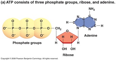
  • ATP- (Adenosine Triphosphate) – This is what we would call the main energy or “money source” of the cell. It has a large amount of energy in storage and it also transports that energy in the cell.
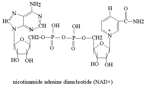
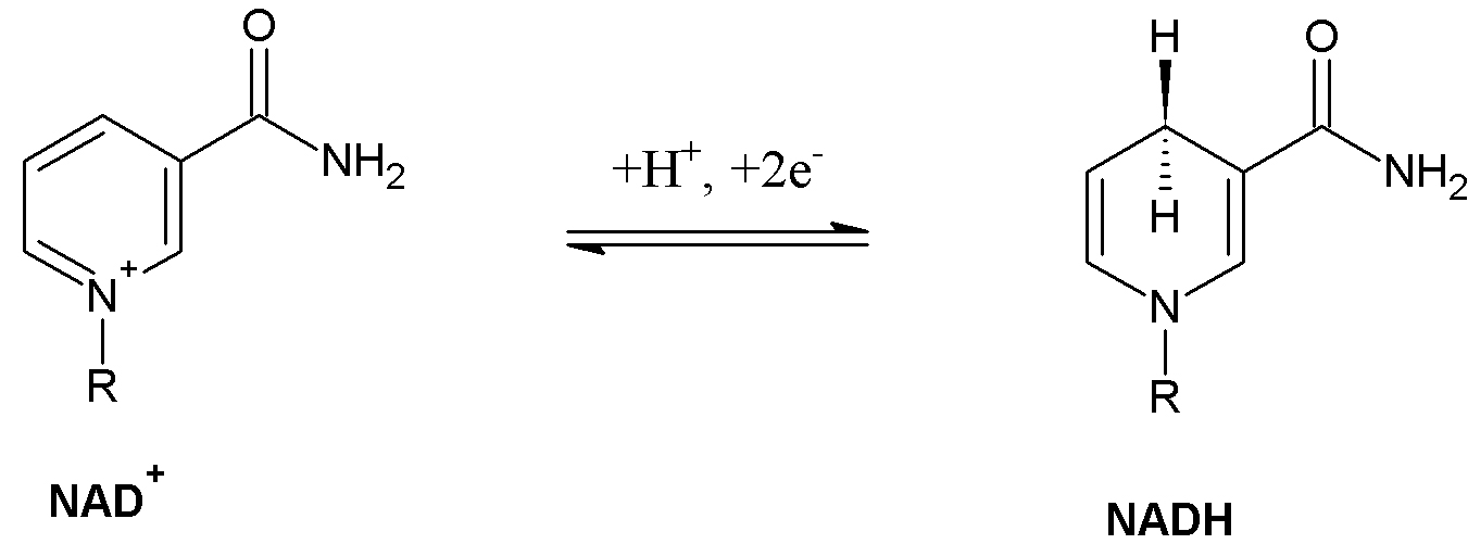
  • NADH An electron carrier with high amounts of energy. It is what transports the electrons made in Glycolysis and Kreb’s cycle to the Electron Transport Chain. In other words, “The Shuttle service.”
  • FADH2 Also an electron carrier with a high amount of energy, this is also a “Shuttle Service.”

Cellular respiration takes place in three main steps:

  1. GLYCOLYSIS

This is when the glucose molecule from food sources is divided into two molecules known as pyruvate. Two ATP molecules are made and also 2 NADH molecules. So for every 1 glucose molecule, we get pyruvate, 2 ATP and 2 NADH.


2. KREB’S CYCLE

Here the pyruvate molecule made in glcolysis is use to 2 ATP and some FADH2 and NADH are also made for the 3rd step.


3. ELECTRON TRANSPORT CHAIN

The NADH and FADH2 made previously are used to form a proton gradient which eventually forms 32 molecules of ATP.


Now that we have recapped what cellular respiration entails, we can now make the link between chemiosmosis and the generation of ATP in the mitochondria.


In the mitochondria of cells, there is something we would call a proton gradient, which is the inner membrane of the mitochondria.

  Electrons are journeying through the electron transport chain, hydrogen ions are pushed or pumped out from the mitochondria into a space known as the inter-membrane space.

 When this happens, a concentration gradient is formed or a more in depth term would be “proton motive force.”

Proton motive force is simply the energy that causes the movement of protons or the “driving force” of protons. 

This causes the protons to move from an area of high concentration to one of lower concentration by diffusion, which in turn, causes the ion to be diffused back into the cell by a process called ATP synthase.

ATP synthase is a form of enzyme that produces ATP by chemiosmosis and it is what allows the protons to be able to go through the membrane.

So to recap: ATP is made in cells that respire by an electrochemical gradient that is found across the inner membrane of the mitochondria. It uses the NADH and FADH2 as the energy source.

Now that the protons have moved back into the membrane by ATP synthase, this same energy that does this, supplies a sufficient amount of energy for ADP to join together with inorganic phosphate to thus form ATP (oxidative phosphorylation).

 This is what it mainly looks like………………………………………………..

Image 101

References for Images:


http://blog.cholesterol-and-health.com/2012/01/we-really-can-make-glucose-from-fatty.html

http://guweb2.gonzaga.edu/faculty/cronk/CHEM440pub/L30-index.cfm

http://apps.cmsfq.edu.ec/biologyexploringlife/text/chapter7/concept7.4.html

http://en.wikipedia.org/wiki/Electron_transport_chain


Video

Glycolysis Made Easy

YouTube Video 2- Glycolysis

This video on glycolysis is very concise and appealing to the eyes. The video makes use of a step by step diagram which is easy to follow. The notes are short and to the point. According to the video glucose is converted to glucose-6-phosphate by adding a phosphate group from ATP in the presence of hexokinase. Glucose-6-phosphate is converted to fructose-6-phosphate by phospoglucose isomerase which is then converted to fructose-1,6-bisphosphate by adding a phosphate group from ATP in the presence of phosphofructokinase. The fructose-1,6-bisphosphate is converted to 2 triose phosphate isomers, dihydroxyacetone Phosphate and glyceraldehyde-3-phosphate, by the enzyme aldose. The enzyme triose phosphate isomerase regulates these isomers by favouring one over the other depending on the concentration of ATP and ADP. When the body requires ATP glceraldehyde-3-phosphate is favoured and is converted to two 1,3-bisphosphoglycerate by the removal of H+ and addition of a phosphate group from NAD+P in the presence of glyceraldehyde phosphate dehydrogenase. 2 ATP is produced by removing a phosphate group in the presence of phosphoglycerate kinase to produce two 3-phosphoglycerate. These are converted to 2-phosphoglycerate by phosphoglyceromutase. Enolase is used to remove 2 water molecules to produce two phosphoenolpyruvate. Finally pyruvate kinase converts the phosphoenolpyruvate to 2 pyruvate by removing a phosphate group per compound to produce 2 ATP. Therefore in total 4 ATP is produced per glucose, 2 ATP is used per glucose, 2 pyruvates is produced per glucose, 2 NADH+H is produced per glucose and 2 water molecules is produced per glucose.

GLYCOLYSIS 1

Hellloooo and Welcome

Our topic for today isssssssssss…………………….

GLYCOLYSIS!!!!!!!!


 One Does Not Simply learn glycolysis - One Does Not Simply learn glycolysis  Boromir

 IMAGE 89

When you hear the word glycolysis ….what comes to mind????

Well lets split it up

Glycoisn’t that like glucose

And

Lysisdoesn’t this mean splitting

So lets put it together …

The splitting of glucose

In this process we begin with the 6 carbon glucose and end with 2 molecules of 3 carbon pyruvate.

There are ten reactions which can be divided into two main stages.

ENERGY INVESTMENT STAGE & ENERGY GENERATION STAGE

In these reactions 10 enzymes are involved…

FIRST STAGE

ENERGY INVESTMENT STAGE

  1. Here glucose is converted into glucose -6 phosphate. ATP is converted into ADP. The enzyme involved is hexokinase . This is a transferase enzyme that transfer a terminal phosphoryl  group from the ATP unto the glucose. This is significant as by the addition of the phosphate group to the glucose, it activates the glucose resulting in it being unstable and this promotes the reaction. This reaction is not reversible however.

 2. Then glucose-6-phosphate is converted into fructose -6-phosphate. The enzyme that catalyses this reaction is phosphohexose isomerase.  This is important as an aldose sugar is being converted to a ketose sugar. This reaction is reversible.

 3. After the fructose 6-phosphate is converted into fructose1,6-bisphosphate. The enzyme responsible for this reaction is phosphofructokinase-1. This enzyme is the most regulated in glycolysis .  In this reaction ATP is converted into ADP. This reaction is not reversible.

 4. Next the fructose 1,6- bisphosphate a 6 carbon compound  is converted into glyceraldehyde 3-phosphate and Dihydroxyacetone  phosphate  both of which are 3 carbon compounds. The enzyme here is aldolase. The reaction is reversible.

 5. Here  the enzyme, triose phosphate isomerase converts the Dihydroxyacetone phosphate into glyceraldehyde 3-phosphate.At the end of this stage there are two glyceraldehyde 3-phosphate molecules.

The Second Stage

ENERGY GENERATION  STAGE

 6. Then the two glyceraldehyde 3-phosphate is converted into two  1,3- bisphosphoglycerate .  Here oxidation and phosphorylation takes place.  Oxidation  is an energetically feasible reaction. NAD+  has gained  a hydrogen. Glyceraldehyde 3-phosphate has lost a hydrogen  and given it to the NAD + . The enzyme is glyceraldehyde 3-phosphate dehydrogenase. This reaction is the only oxidation reaction of glycolysis. This reaction is reversible.


7. The two 1,3- bisphosphoglycerate  is then converted into two  3-phosphoglycerate. The enzyme that catalyses this reaction is phosphoglycerate kinase. It is a reversible reaction. Here two molecules of ADP is converted to two molecules of ATP. This is the first reaction that produces ATP.

8.  Here the two molecules of 3- phosphoglycerate is converted to two molecules of 2- phosphoglycerate. The enzyme for this reaction is phosphoglycerate mutase. Firstly the 3- phosphoglycerate is converted into an intermediate called 2,3- bisphosphoglycerate and then the enzyme phosphoglycerate mutase removes the  phosphate group on carbon 3 to  end up with two molecules of 2- phosphoglycerate.This reaction is reversible

 9. The two molecules of  2-phosphoglycerate is converted to two molecules of phosphoenol pyruvate. The enzyme for this step is enolase. There is the loss of water in this step or a dehydration reaction. The phosphoenol pyruvate has alot of energy. This reaction is reversible.

 10. The two molecules of phophoenol pyruvate  is then converted to two molecules of pyruvate . The enzyme here is pyruvate kinase   .This reaction is the most energetically favourable in glycolysis and it is not reversible.


Image 90

So folks we have went through each step and I am sure we have understood them.

Hope this has enlightened you on the topic glycolysis…….

See you again soooooooooooon……………….


Image 91

 Reference for Images:

http://quotespics.com/one-does-not-simply-learn-chinese-lord-of-the-rings/

http://www.lactate.com/triathlon/lactate_triathlon_anaerobic_energy.html

http://funny-pics.co/wp-content/uploads/funny-animal-bye-bye-445×299.jpg